

SOME METHODS USED IN HLA-B27 DIAGNOSTIC TESTING
Since 1973, the strong association between the human leukocyte antigen HLA-B27 and ankylosing spondylitis (AS) has been demonstrated.1 HLA-B27 is associated with promoting early disease progression, more severe arthritis, and the appearance of extra-articular features. Many studies have shown that the gene encoding this protein is present in about 90% of patients with AS and in about 50-80% of patients with other spondyloarthritis-related diseases. 2, 3
According to the "Guidelines for the Diagnosis and Treatment of Musculoskeletal Diseases" issued by the Ministry of Health (MOH) with Decision No. 361/QD-BYT, HLA-B27 testing is one of the paraclinical tests that support the diagnosis of AS. Diagnosing the disease at the right time and promptly is key to managing musculoskeletal diseases and leads to a better treatment prognosis.4 In addition, HLA-B27 test results also help in selecting drugs to treat certain autoimmune diseases and in determining the risk of developing adverse drug reactions. Currently, the techniques used to detect HLA-B27 include microcytotoxicity, Flow Cytometry, Real-time PCR, PCR-SSP, Microarray, sequencing, etc., but the most common are still Flow Cytometry, PCR-SSP, and Real-time PCR, each with its own advantages. 5, 6
Flow Cytometry (Flow-Based Cell Analysis)
Flow Cytometry is a tool widely applied in many fields such as immunology, virology, molecular biology, cancer biology, etc. This method allows the analysis of multiple parameters of single cells or particles. The basic measurement principle involves light scattering and fluorescence emission, which occur when light from an excitation source (usually a laser beam) strikes particles moving in a solution. The data obtained can provide valuable information on biochemical, biophysical, and molecular aspects. The ability to scatter light or bind fluorescent dyes depends on the size, internal structure, and surface composition of the cell. 7, 8
The HLA-B27 test uses fluorescently labeled anti-HLA-B27 monoclonal antibodies that bind to this antigen (if present) on the surface of white blood cells in the patient sample (Figure 1). The sample is then analyzed by the instrument system and data processing software to detect the presence of the HLA-B27 antigen.5
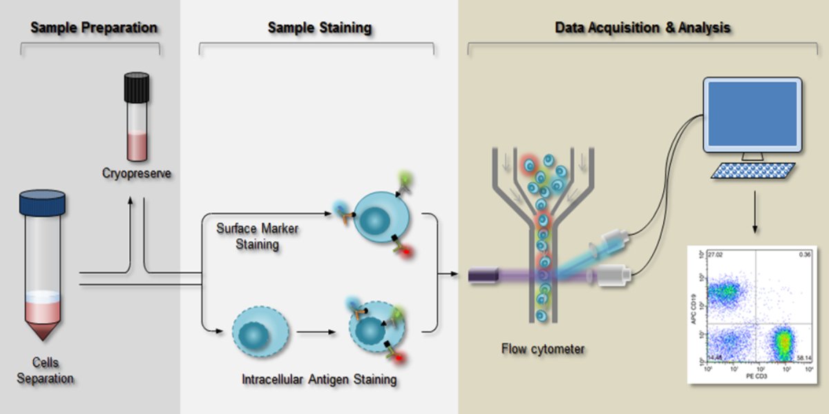
Figure 1: Flow Cytometry workflow9
Flow Cytometry has many advantages, such as fast turnaround, high throughput, and relatively low cost, but its specificity is lower than that of PCR techniques. This is due to the presence of other HLA antigens that cross-react with HLA-B27 and lead to false-positive results, especially HLA-B7 and HLA-B37. Different anti-HLA-B27 antibody clones have different affinities for these antigens. Some studies indicate that, to increase reliability, at least two HLA-B27 monoclonal antibodies need to be used in the test. For some inconclusive results, other methods such as PCR or sequencing are needed for confirmation. 10
PCR-SSP (Sequence-Specific Primer Polymerase Chain Reaction)
Besides Flow Cytometry, PCR-based methods are also widely used in clinical practice. PCR-SSP (Polymerase Chain Reaction-Sequence Specific Primers) is a form of PCR, a DNA amplification method based on thermal cycles. First introduced in the early 1990s, the PCR-SSP technique is based on the principle that primers perfectly matched to the template strand give better amplification efficiency than mismatched primers. Specificity is determined by the use of sequence-specific primers, in which a mismatch at the 3' terminal nucleotide inhibits the initiation of a nonspecific reaction, because Taq polymerase lacks 3'-5' exonuclease activity. Therefore, only the desired allele is selectively amplified, and the product can be detected by agarose gel electrophoresis. This technique is applied in many tests, such as identifying single nucleotide point mutations, blood group genotyping, HLA typing, etc. 11, 12
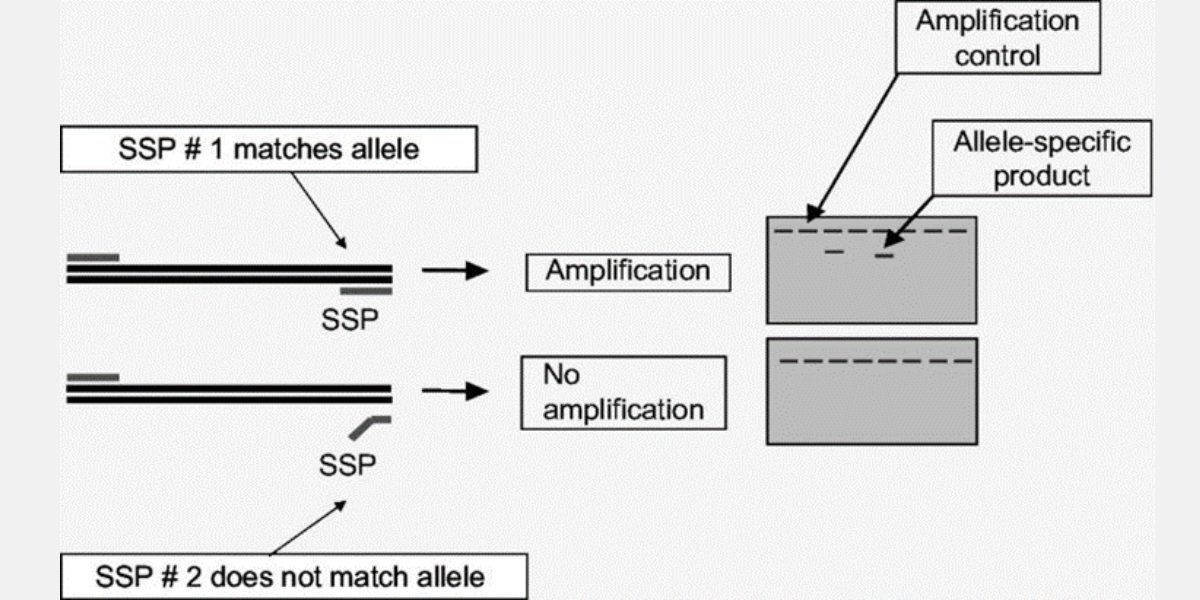
Figure 2: Principle of the SSP-PCR technique 13
In HLA-B27 testing, the genomic DNA sample obtained after extraction is amplified with primers specific to the HLA-B27 allele. After analysis by agarose gel electrophoresis, the appearance of a band of the expected size indicates the presence of this allele in the sample. In addition, the PCR-SSP technique is also used to determine HLA-B27 subtypes.14
With the advantages of high sensitivity, high specificity, and low cost, PCR-SSP is commonly used in laboratory settings. These techniques still have some limitations, such as the inability to detect homozygous HLA-B27 samples, or the need to reopen the product tubes after PCR for electrophoresis, which increases the risk of contamination.14
Real-time PCR (Real-Time Polymerase Chain Reaction)
In Real-time PCR, amplification products are detected using fluorescent dyes. These dyes are attached to oligonucleotide probes that bind specifically to the amplification products. During the extension phase, the exonuclease activity of the enzyme degrades the fluorescent probe and releases the fluorescent dye from its proximity to the quencher. The fluorescence signal increases after each cycle if amplification occurs and is measured at the end of each qPCR cycle. Real-time monitoring of fluorescence intensity allows detection and quantification of the accumulated product. Current applications of Real-time PCR include gene expression analysis, mutation detection, pathogen detection and quantification, detection of genetically modified organisms, allergen detection, etc. 15, 16
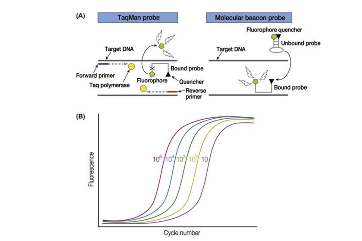
Figure 3: Principle of Real-time PCR 16
To detect the gene encoding HLA-B27, primers are usually designed to be specific to exon 2 or exon 3 of the gene. The test includes primers for a housekeeping gene such as beta-globin to control for the presence of cellular material in the sample. Test results are calculated based on Ct values without reopening the tubes after the reaction. This overcomes a limitation of conventional PCR methods. In addition, Real-time PCR has high sensitivity and specificity, helping to deliver accurate test results quickly and to confirm difficult cases.17
Our Solution:
VIET HUY Technological Scientific Equipment Co., Ltd currently supplies the HLA-B27 REAL-TM kit, by Sacace Biotechnologies - Italy. The product is now used in many hospitals and clinics across the country.
References:
1. Brewerton DA, Hart FD, Nicholls A, Caffrey M, James DC, Sturrock RD. Ankylosing spondylitis and HL-A 27. Lancet. 1973 Apr 28;1(7809):904-7. doi: 10.1016/s0140-6736(73)91360-3. PMID: 4123836.
2. Marc C. Hochberg, Alan J. Silman, Josef S. Smolen, Michael E. Weinblatt, Michael H. Weisman. Rheumatology (Sixth Edition). Mosby. 2015. Page iv. ISBN 9780323091381. https://doi.org/10.1016/B978-0-323-09138-1.00251-5.
3. van Vollenhoven, Ronald. (2017). Evaluation and Differential Diagnosis of Polyarthritis. 10.1016/B978-0-323-31696-5.00042-5.
4. Guidelines for the Diagnosis and Treatment of Musculoskeletal Diseases [In Vietnamese] (Issued together with Decision No. 361/QD-BYT dated Jan 25, 2014, Minister of Health).
5. Chheda P et al. HLA-B27 testing: A journey from flow cytometry to molecular subtyping. J Clin Lab Anal. 2018 Jun;32(5):e22382.
6. Parasannanavar DJ, Rajadhyaksha A, Ghosh K. Application of a Simple In-House PCR-SSP Technique for HLA-B* 27 Typing in Spondyloarthritis Patients. Arthritis. 2013;2013:504109. doi: 10.1155/2013/504109. Epub 2013 Dec 19. PMID: 24490069; PMCID: PMC3880732.
7. McKinnon KM. Flow Cytometry: An Overview. Curr Protoc Immunol. 2018 Feb 21;120:5.1.1-5.1.11. doi: 10.1002/cpim.40. PMID: 29512141; PMCID: PMC5939936.
8. Aysun Adan, Günel Alizada, Yağmur Kiraz, Yusuf Baran & Ayten Nalbant (2016): Flow cytometry: basic principles and applications, Critical Reviews in Biotechnology, DOI: 10.3109/07388551.2015.1128876
9. F. Balny et at. 70e Congres de l'AACC 2018 (American Association for Clinical Chemistry), IRBM News, Volume 40, Issue 1, 2019, Pages 1-33, ISSN 1959-7568,https://doi.org/10.1016/j.irbmnw.2018.12.001.
10. Sandor Barath et al. Combined use of different antibody clones improves the efficiency of human leukocyte antigen B27 detection by flow cytometry. (2020). https://doi.org/10.1002/cyto.b.21989.
11. Welsh K et al. Molecular typing for the MHC with PCR-SSP. Rev Immunogenet. 1999;1(2):157-76. PMID: 11253945.
12. Musa et al. A simple PCR-SSP method for detection of HLA-B*15:02, *15:13, and *15:21. Turkish Journal of Biochemistry, vol. 48, no. 3, 2023, pp. 230-233. https://doi.org/10.1515/tjb-2022-0202.
13. Marino et al. (2008). The Human Major Histocompatibility Complex and DNA- Based Typing of Human Leukocyte Antigens for Transplantation.
14. Lara-Armi FF et al. Optimization of HLA-B*27 ALLELE Genotyping by PCR-SSP. Clinics (Sao Paulo). 2020 Oct 26;75:e1840. doi: 10.6061/clinics/2020/e1840. PMID: 33146354; PMCID: PMC7561065.
15. Artika IM et al. Real-Time Polymerase Chain Reaction: Current Techniques, Applications, and Role in COVID-19 Diagnosis. Genes (Basel). 2022 Dec 16;13(12):2387. doi: 10.3390/genes13122387. PMID: 36553654; PMCID: PMC9778061.
16. Wang-Shick Ryu, Chapter 4 - Diagnosis and Methods, Molecular Virology of Human Pathogenic Viruses, Academic Press, 2017, Pages 47-62, ISBN 9780128008386, https://doi.org/10.1016/B978-0-12-800838-6.00004-7.
Related Articles

GENEQUALITY 2050 - A COMPREHENSIVE AUTOMATION SOLUTION FROM AB ANALITICA
The GeneQuality 2050 system - a comprehensive automation solution from AB Analitica, delivering outstanding performance in real-time PCR testing with speed, accuracy, and safety.

Next-Generation Sequencing (NGS) in BRCA Mutation Analysis: The Optimal Tool for Diagnosing Hereditary Breast and Ovarian Cancer
Next-generation sequencing (NGS) helps detect BRCA mutations in hereditary breast and ovarian cancer, delivering high accuracy and improving treatment approaches.

GENEQUALITY Whole Exome Sequencing (WES) | Viet Huy
GENEQUALITY Whole Exome Sequencing (WES) is a whole exome sequencing solution developed by AB ANALITICA and CE-IVD certified under EU Regulation 2017/746 (IVDR). This product complies...
Need Advice on the Right Testing Solution?
Viet Huy engineers are ready to help your laboratory choose the optimal technology.